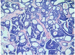
Il carcinoma cribriforme è così chiamato perché al microscopio ottico si osservano degli spazi vuoti tra le cellule che lo fanno assomigliare ad un setaccio. Spesso il carcinoma cribriforme è associato al carcinoma duttale in situ (DCIS) (1-4). Presenta un’incidenza del 0.3-4% fra tutte le neoplasie maligne mammarie primarie.
Diagnosi:
- Palpazione: il ca. cribriforme invasivo (ICC) si apprezza come un piccolo (2,5 cm) nodulo o un’area che sembra più consistente rispetto al resto parenchima mammario.
- Mammografia
- USG
- Biopsia
- Aspirazione con ago sottile (FNA)
Esame Prognosi: buona a motivo della lenta crescita.
Esame anatomo-patologico: La massa del tumore è solida, di colorito grigio-bianca, dura, acapsulata. Se il tumore ha un pattern invasivo e cribriforme >90% ed è associato a meno del 50% di una componente tubulare, allora dovrebbe essere classificato come un ICC classico. Tuttavia, se il tumore ha un pattern invasivo e un pattern cribriforme, ma è accompagnato dal 10% al 49% di altre componenti morfologiche (escluso il carcinoma tubulare), allora dovrebbe essere classificato come ICC misto.
Terapia chirurgica in prima istanza: prevede lumpectomia o mastectomia in base alle dimensioni della neoplasia e se è interessata più di un’area della mammella.
Terapia adiuvante include:
- radioterapia
- terapia ormonale: la maggior parte dei tumori al seno cribriformi sono ER positivi (9).
- chemioterapia
- terapia mirata (target therapy)
- bifosfonati
- Chao-Hua Mo et al: Invasive cribriform carcinoma of the breast: a clinicopathological analysis of 12 cases with review of literature. Int J Clin Exp Pathol. 2017; 10(9): 9917–9924.
- Venable JG, Schwartz AM, Silverberg SG. Infiltrating cribriform carcinoma of the breast: a distinctive clinicopathologic entity. Hum Pathol. 1990;21:333–338.
- Marzullo F, Zito FA, Marzullo A, Labriola A, Schittulli F, Gargano G, De Girolamo R, Colonna F. Infiltrating cribriform carcinoma of the breast. A clinico-pathologic and immunohistochemical study of 5 cases. Eur J Gynaecol Oncol. 1996;17:228–231.
- Zhang W, Zhang T, Lin Z, Zhang X, Liu F, Wang Y, Liu H, Yang Y, Niu Y. Invasive cribriform carcinoma in a Chinese population: comparison with low-grade invasive ductal carcinomanot otherwise specified. Int J Clin Exp Pathol. 2013;6:445–457.
- Rakha E, Pinder SE, Shin SJ, Tsuda H. Tubular carcinoma and cribriform carcinoma. In: Lakhani SR, Ellis IO, Schnitt SJ, Tan PH, van de Vijver MJ, editors. WHO classification of tumours of the breast. 4th edition. Lyon: IARC Press; 2012. pp. 43–45.
- Liu XY, Jiang YZ, Liu YR, Zuo WJ, Shao ZM. Clinicopathological characteristics and survival outcomes of invasive cribriform carcinoma of breast: a SEER population-based study. Medicine (Baltimore) 2015;94:e1309. [PMC free article] [PubMed] [Google Scholar]
- Zhang W, Lin Z, Zhang T, Liu F, Niu Y. A pure invasive cribriform carcinoma of the breast with bone metastasis if untreated for thirteen years: a case report and literature review. World J Surg Oncol. 2012;10:251.
- Lee YJ, Choi BB, Suh KS. Invasive cribriform carcinoma of the breast: mammographic, sonographic, MRI, and 18 F-FDG PET-CT features. Acta Radiol. 2015;56:644–651.
- Colleoni M, Rotmensz N, Maisonneuve P, Mastropasqua MG, Luini A, Veronesi P, Intra M, Montagna E, Cancello G, Cardillo A, Mazza M, Perri G, Iorfida M, Pruneri G, Goldhirsch A, Viale G. Outcome of special types of luminal breast cancer. Ann Oncol. 2012;23:1428–1436.
- Cong Y, Qiao G, Zou H, Lin J, Wang X, Li X, Li Y, Zhu S. Invasive cribriform carcinoma of the breast: a report of nine cases and a review of the literature. Oncol Lett. 2015;9:1753–1758.
- Nishimura R, Ohsumi S, Teramoto N, Yamakawa T, Saeki T, Takashima S. Invasive cribriform carcinoma with extensive microcalcifications in the male breast. Breast Cancer. 2005;12:145–148.
- Stutz JA, Evans AJ, Pinder S, Ellis IO, Yeoman LJ, Wilson AR, Sibbering DM. The radiological appearances of invasive cribriform carcinoma of the breast. Nottingham Breast Team. Clin Radiol. 1994;49:693–695. [PubMed] [Google Scholar]
- Lim HS, Jeong SJ, Lee JS, Park MH, Yoon JH, Kim JW, Park JG, Kang HK. Sonographic findings of invasive cribriform carcinoma of the breast. J Ultrasound Med. 2011;30:701–705.
- Shousha S, Schoenfeld A, Moss J, Shore I, Sinnett HD. Light and electron microscopic study of an invasive cribriform carcinoma with extensive microcalcification developing in a breast with silicone augmentation. Ultrastruct Pathol. 1994;18:519–523.
- Puri S, Mohindroo S, Gulati A. Collagenous spherulosis: an interesting cytological finding in breast lesion. Cytojournal. 2015;12:25.
- Azoulay S, Lae M, Freneaux P, Merle S, Al Ghuzlan A, Chnecker C, Rosty C, Klijanienko J, Sigal-Zafrani B, Salmon R, Fourquet A, Sastre-Garau X, Vincent-Salomon A. KIT is highly expressed in adenoid cystic carcinoma of the breast, a basal-like carcinoma associated with a favorable outcome. Mod Pathol. 2005;18:1623–1631.
- Arps DP, Chan MP, Patel RM, Andea AA. Primary cutaneous cribriform carcinoma: report of six cases with clinicopathologic data and immunohistochemical profile. J Cutan Pathol. 2015;42:379–387.
- Dobruch-Sobczak K, Roszkowska-Purska K, Chrapowicki E. Cribriform carcinoma mimicking breast abscess – case report. Diagnostic and therapeutic management. J Ultrason. 2013;13:222–229.


